Hamstring strains are common, recur often, and player unavailability is costly. Early, targeted insight helps keep athletes on the pitch.
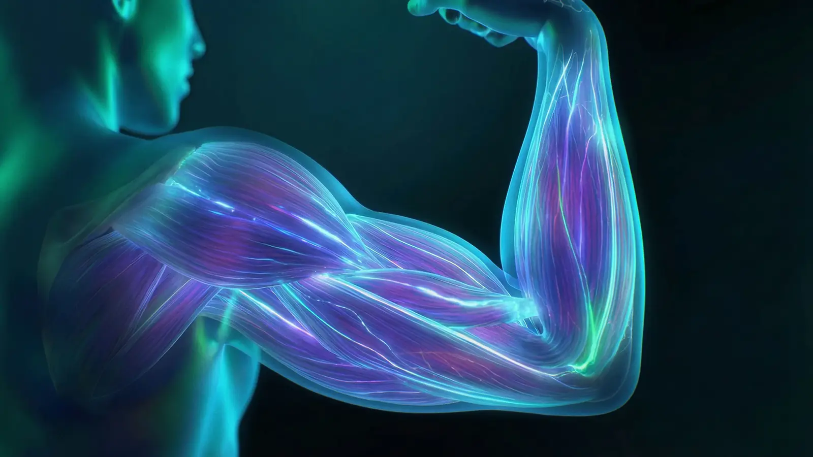
Beyond jump and strength outputs, EMG shows which hamstrings are being recruited within each exercise, exposing asymmetries and compensations.
This hamstring screening revealed greater contribution from the right hamstrings and a left biceps femoris and left semitendinosus asymmetry, providing crucial information to guide programme design, exercise selection, and technique optimisation.
Hamstring injuries remain a major concern affecting athletes in running sports, accounting for approximately 10% of all injuries in field sports [1]. Within the hamstring group, the long head of the biceps femoris is the most frequently injured muscle [2, 3]. During sprinting, the hamstrings extend the hip and flex the knee, but in the late swing phase they shift to an eccentric role, decelerating the forward motion of the leg just before ground contact [4]. It is in this phase, when the muscles are both highly activated, contracting eccentrically and stretched to long lengths, that hamstring injuries typically occur.
Post-injury, changes in biomechanics are common as the neuromuscular system adapts sprint mechanics, such as shorter stride length and reduced hip extension, to protect the previously injured muscle [5]. While these adjustments allow the movement to be completed successfully, they often come at the expense of running efficiency and can increase the load on other hamstring and gluteal muscles. Over time, this compensatory strategy may elevate the risk of reinjury.
The tendency of hamstring injuries to recur, typically around 15-30% [6], with rates as high as 63% reported in some studies [7], is equally troubling. With the frequency and intensity of sport seemingly ever increasing, and player unavailability costly for clubs, the need for better tools to monitor, screen, and understand athletes’ neuromuscular function becomes more pressing. Electromyography (EMG) is one tool that can provide additional insights into this area by investigating these asymmetries, compensations and more.
In applied sport, jump and strength tests are often used to monitor rehabilitation after injury. While these tests provide useful values, they can’t reveal how those results were achieved. Compensations following hamstring injury mean that similar performance values can be achieved through different activation strategies. EMG exposes these compensations, such as asymmetries in activation between medial and lateral hamstrings, which may increase reinjury risk. By combining EMG with other metrics, such as force, we can build a clearer picture of which muscles are producing actively recruited, whether asymmetries or inefficiencies are present, and ultimately, how these factors may influence injury or reinjury risk.
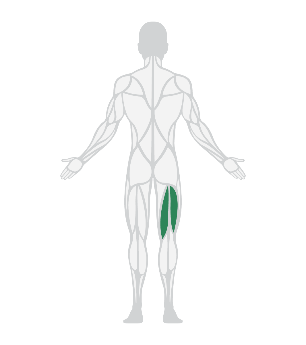
By measuring activity from the biceps femoris and semitendinosus on both legs, practitioners can see the relative contributions of each muscle during specific exercises. When used in this context, EMG helps practitioners use common exercises as a screening tool, revealing which muscles are most optimally recruited and what needs to change to allow an athlete to progress or reduce injury risk.
In this blog we examined muscle activation during six common hamstring exercises, using EMG to assess bilateral biceps femoris and semitendinosus contribution.
One healthy male participant completed a range of hamstring rehabilitation exercises. Four Trigno Avanti sensors were paired with a Trigno Lite receiver, and placed on:
Each location for sensor placement was shaved to remove any excess hair, cleaned with an alcohol wipe and then each Trigno Avanti sensor was placed following the SENIAM guidelines.
After placement, each sensor was normalised to a Maximum Voluntary Contraction (MVC). Normalisation is paramount when collecting surface EMG, to reduce the effect of the limitations of surface EMG, whilst providing a reference value to allow comparisons between muscles, athletes and sessions.
To assess the contribution of the hamstring muscles during different exercises, the following exercises were performed in a randomised order with 2 minutes rest between each exercise:
These exercises were selected due to their frequent usage in hamstring testing and rehabilitation programmes. EMG data was collected through Trigno Discover and all amplitudes normalised to the muscle specific MVC.
Note that dynamic exercises can exceed 100% of isometric MVC.
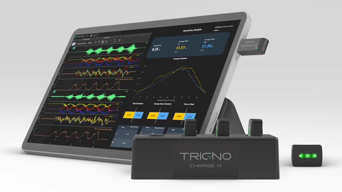
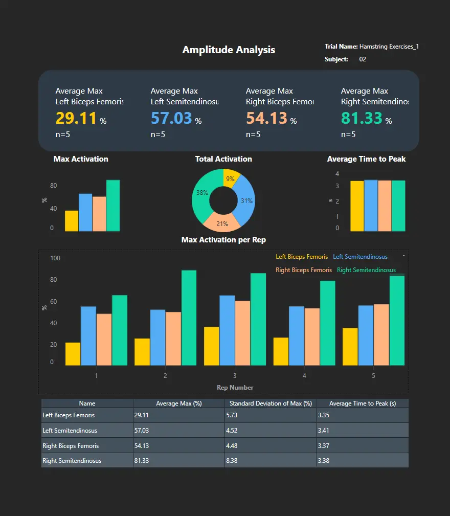
Using the Trigno Analytics feature, reports were generated immediately after each exercise. Across the session, the right hamstrings typically showed higher activation levels than the left, reflected in both higher Average Max (%MVC) and a larger share of Total Activation. The standout finding was the under-recruitment of left BF across most tasks alongside comparatively high activity in the other muscles measured, illustrated clearly by the Amplitude reports for Bridges (Figure 1) and Romanian Deadlifts (Figure 2).
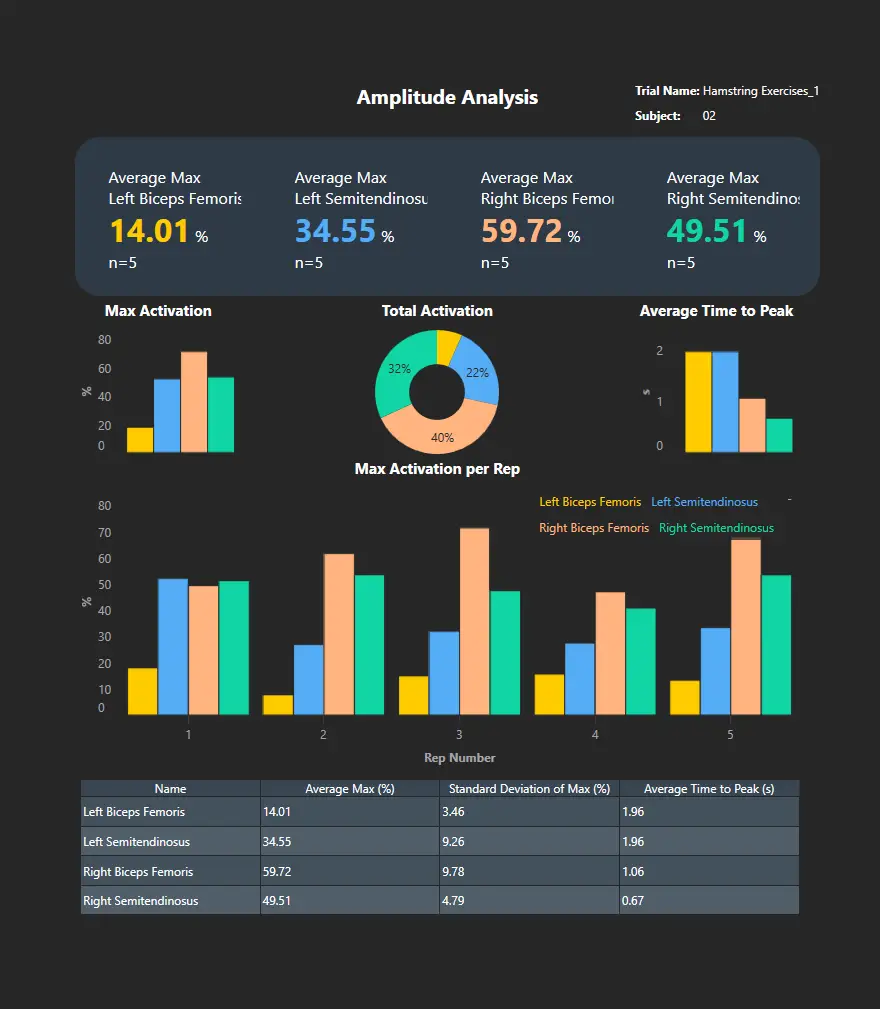
Symmetry reports further highlight the large asymmetry between left BF and left ST activation (Bridges – Figure 3), a common theme throughout the exercises. The only exception to this recruitment pattern was during Nordic Hamstring Curls. Because the left BF does reach high activation in the Nordics, this muscle may be under-recruited during hamstring exercises due to task or technique issues, raising some considerations and questions:
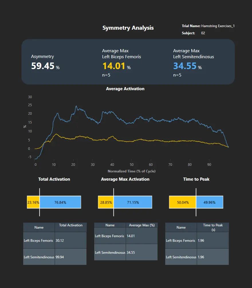
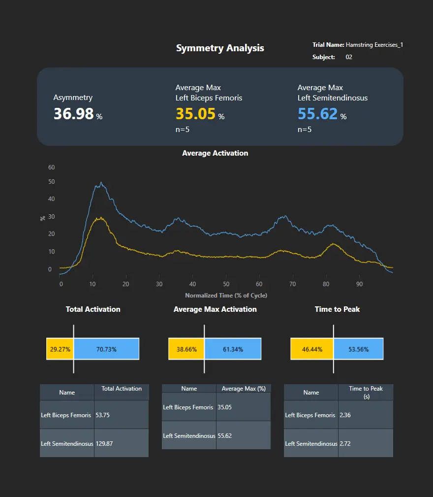
Table 1 – Avg. Max EMG Amplitude (%MVC) for each exercise and each muscle, taken from the Trigno Analytics Amplitude Reports. All colours match those in the reports, with the lowest and highest activation for each muscle underlined and bold respectively.
| Left Biceps Femoris | Left Semitendinosus | Right Biceps Femoris | Right Semitendinosus | |
|---|---|---|---|---|
| Bridges | 14.01% | 34.55% | 59.72% | 49.51% |
| Single Leg Bridges | 35.05% | 55.62% | 76.64% | 68.17% |
| Long Lever Bridges | 13.87% | 66.51% | 56.57% | 58.38% |
| Romanian Deadlift | 29.11% | 57.03% | 54.13% | 81.33% |
| Roman Chair | 35.82% | 93.69% | 100.49% | 113.24% |
| Nordic Hamstring Curls | 139.78% | 146.75% | 152.85% | 128.99% |
EMG adds an extra, crucial layer to athlete monitoring through hamstring screening. It shows how the hamstrings contribute, not just the performance outcome, allowing the right muscle to be targeted with the right exercise and verify changes. EMG complements strength and jump tests rather than replacing them, by giving practitioners the missing neuromuscular context behind those numbers.