Within different disease populations, changes in regional distribution have been seen to signify maladaptation from the associated mechanical or neurological deficit.
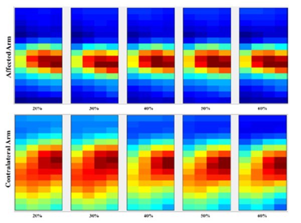
For instance, in stroke populations, studies have shown alterations in muscle architecture including the loss of muscle fibers and overall motor unit number reduction in paretic muscles of stroke survivors. Rasool et al. (2015) (Spatial analysis of muscular activations in stroke survivors – PubMed (nih.gov)) showed regional changes in amplitude clustering when comparing affected and unaffected limbs following a stroke.
The overall shrinkage of the area of activation that was seen in the affected side could likely be a sign of muscle atrophy. While the spatial pattern of the sEMG may refer to the mechanisms of motor unit recruitment within a muscle, the consistency of such patterns across force levels confirms the presence of mechanisms that do not change with the force requirements and hence the potential loss of motor unit numbers from de-innervation or disruption of the descending central nervous system pathways.

Graphic reproduced from Rasool G, Afsharipour B, Suresh NL, Xiaogang Hu, Rymer WZ. Spatial analysis of muscular activations in stroke survivors. Annu Int Conf IEEE Eng Med Biol Soc. 2015;2015:6058-61. doi: 10.1109/EMBC.2015.7319773. PMID: 26737673.
By using HDsEMG, disease progression or rehabilitation outcomes can be measured by assessing the regional distribution of the EMG signal and how this changes in relation to specific interventions.

